Advanced Quantitative Fluorescence Microscopy to Probe the Molecular Dynamics of Viral Entry, Science Lab
Por um escritor misterioso
Descrição
Viral entry into the host cell requires the coordination of many cellular and viral proteins in a precise order. Modern microscopy techniques are now allowing researchers to investigate these interactions with higher spatiotemporal resolution than ever before. Here we present two examples from the field of HIV research that make use of an innovative quantitative imaging approach as well as cutting edge fluorescence lifetime-based confocal microscopy methods to gain novel insights into how HIV fuses to cell membranes and enters the cell.
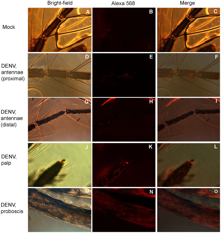
Microscopy in Virology, Science Lab

Single-molecule localization microscopy
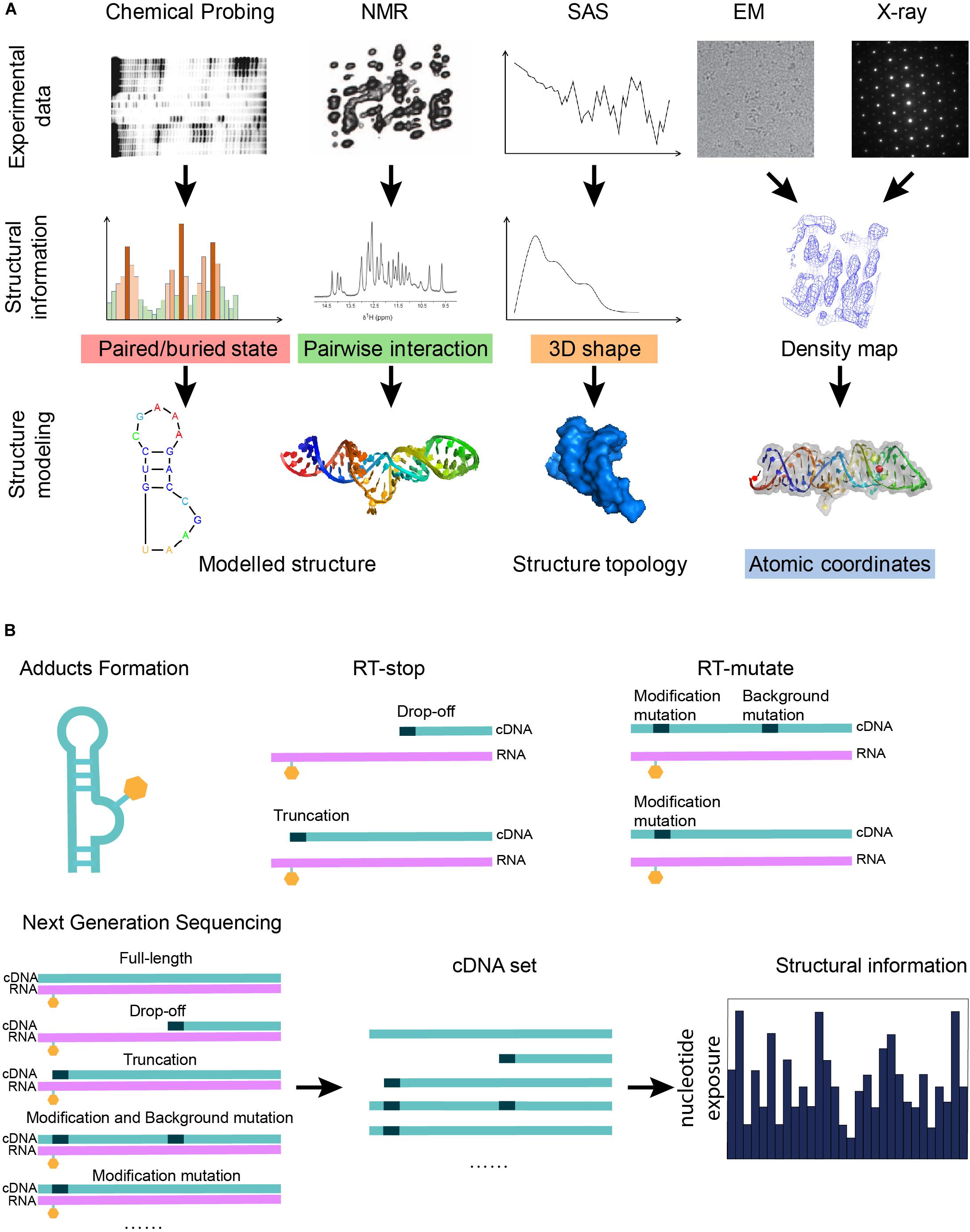
Frontiers Advances in RNA 3D Structure Modeling Using Experimental Data
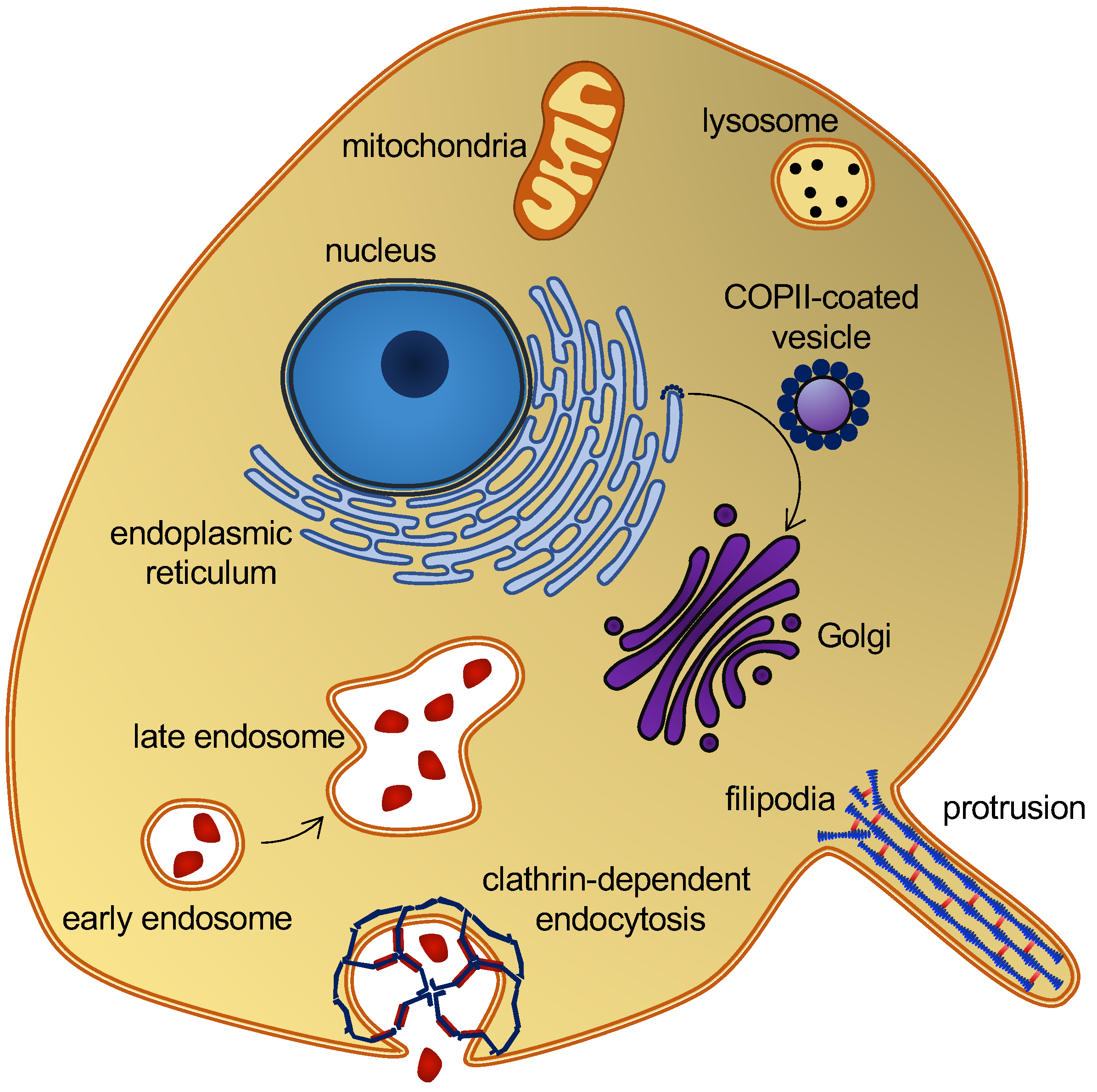
IJMS, Free Full-Text
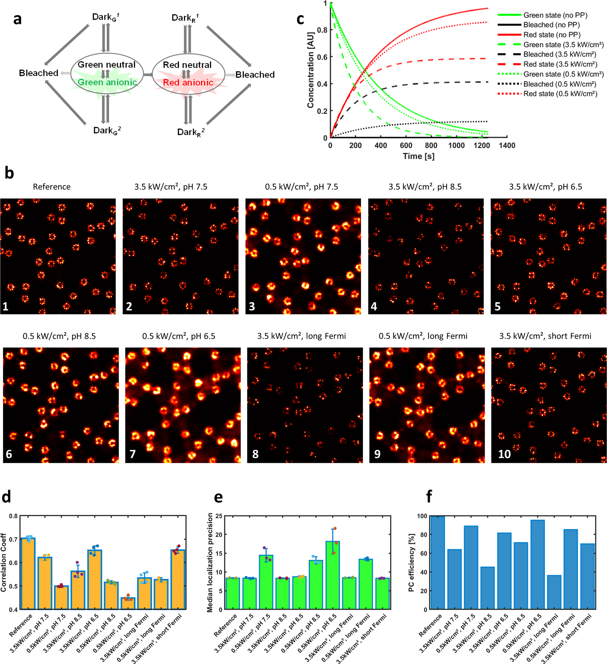
Single molecule imaging simulations with advanced fluorophore photophysics

Ultrafast, sensitive, and portable detection of COVID-19 IgG using flexible organic electrochemical transistors
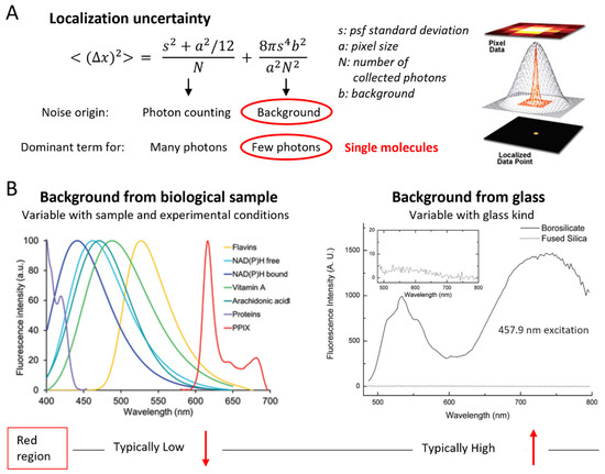
IJMS, Free Full-Text

Mapping of functional SARS-CoV-2 receptors in human lungs establishes differences in variant binding and SLC1A5 as a viral entry modulator of hACE2 - eBioMedicine

Full article: Manipulation of matter with shaped-pulse light field and its applications

Beyond mass spectrometry, the next step in proteomics

CRISPR‐Cas Biochemistry and CRISPR‐Based Molecular Diagnostics - Weng - 2023 - Angewandte Chemie International Edition - Wiley Online Library
Molecular beacons. (a) The most common molecular beacon structure is a
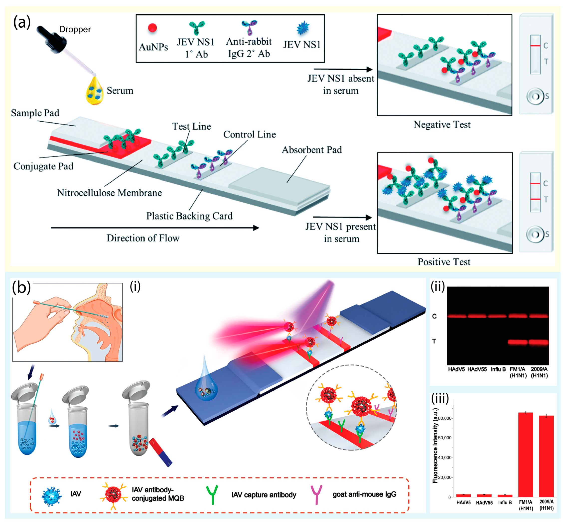
Biosensors, Free Full-Text

Molecular and Immunological Diagnostic Tests of COVID-19: Current Status and Challenges - ScienceDirect

HIV-1 Vpr induces ciTRAN to prevent transcriptional repression of the provirus


/cdn.vox-cdn.com/uploads/chorus_image/image/6264415/valve_booth_ces13.0.jpg)


