Echo image of right ventricular apex (RVA) pacemaker, pressure
Por um escritor misterioso
Descrição

Echo image of right ventricular apex (RVA) pacemaker, pressure
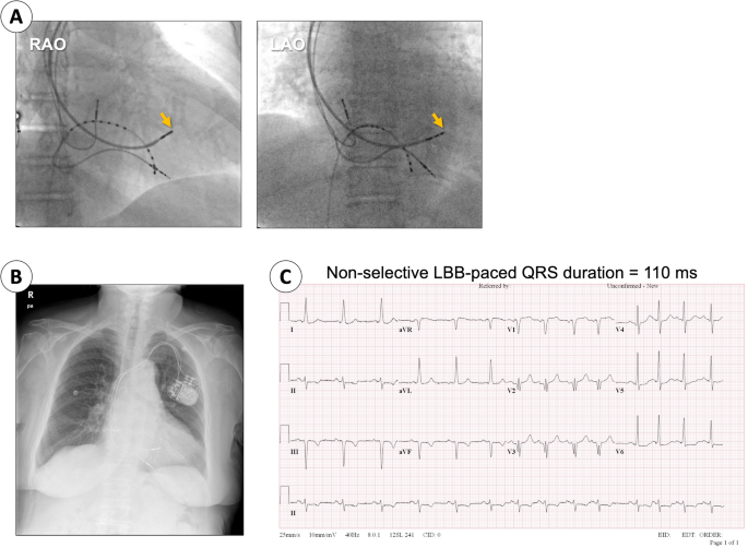
Reversal of pacing-induced cardiomyopathy after left bundle branch area pacing: a case report, International Journal of Arrhythmia

Nonapical Right Ventricular Pacing Is Associated with Less Tricuspid Valve Interference and Long-Term Progress of Tricuspid Regurgitation - ScienceDirect
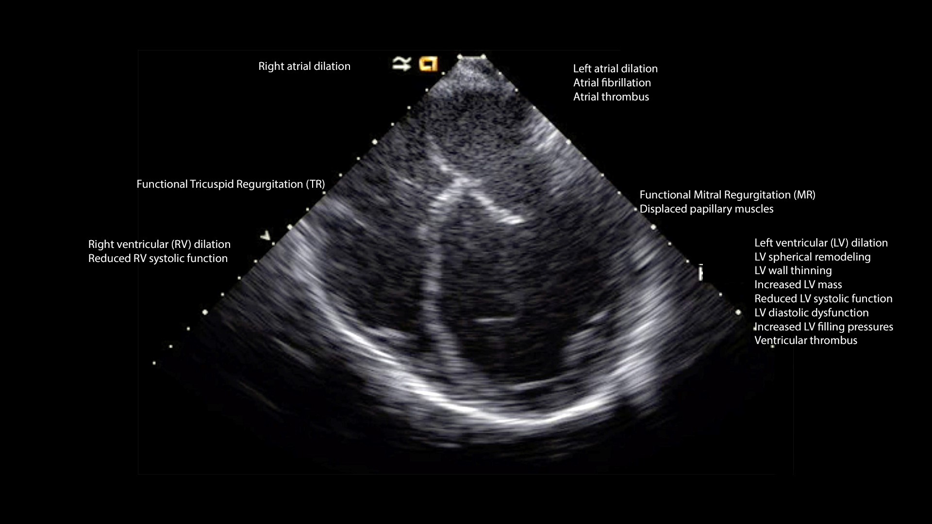
Cardiomyopathy - Congenital Cardiac Anesthesia Society
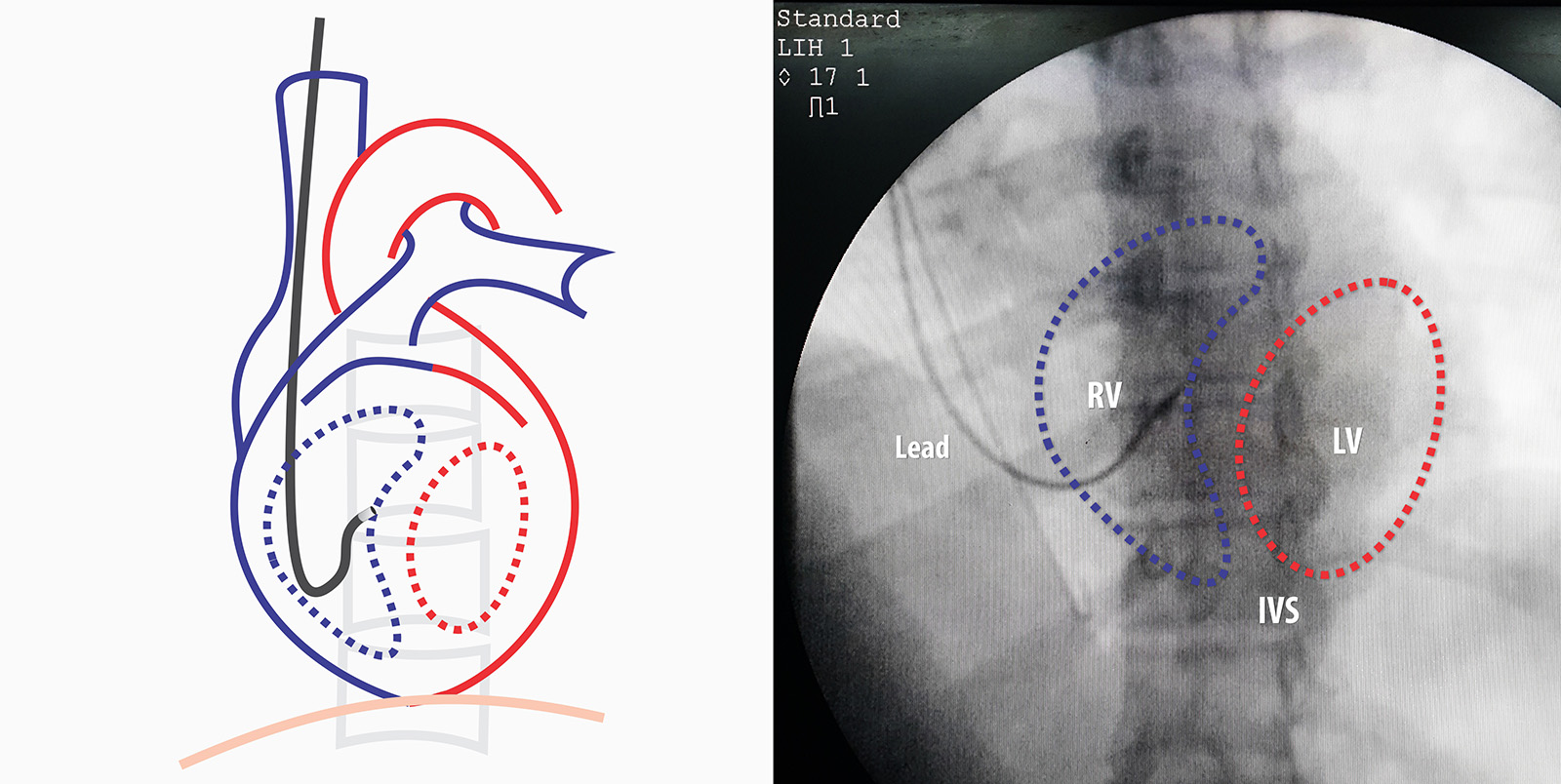
Septal Implantation of Right Ventricular Lead – How to Pace

Adverse effects of right ventricular pacing on cardiac function: prevalence, prevention and treatment with physiologic pacing - ScienceDirect

Advantage of right ventricular outflow tract pacing on cardiac function and coronary circulation in comparison with right ventricular apex pacing.
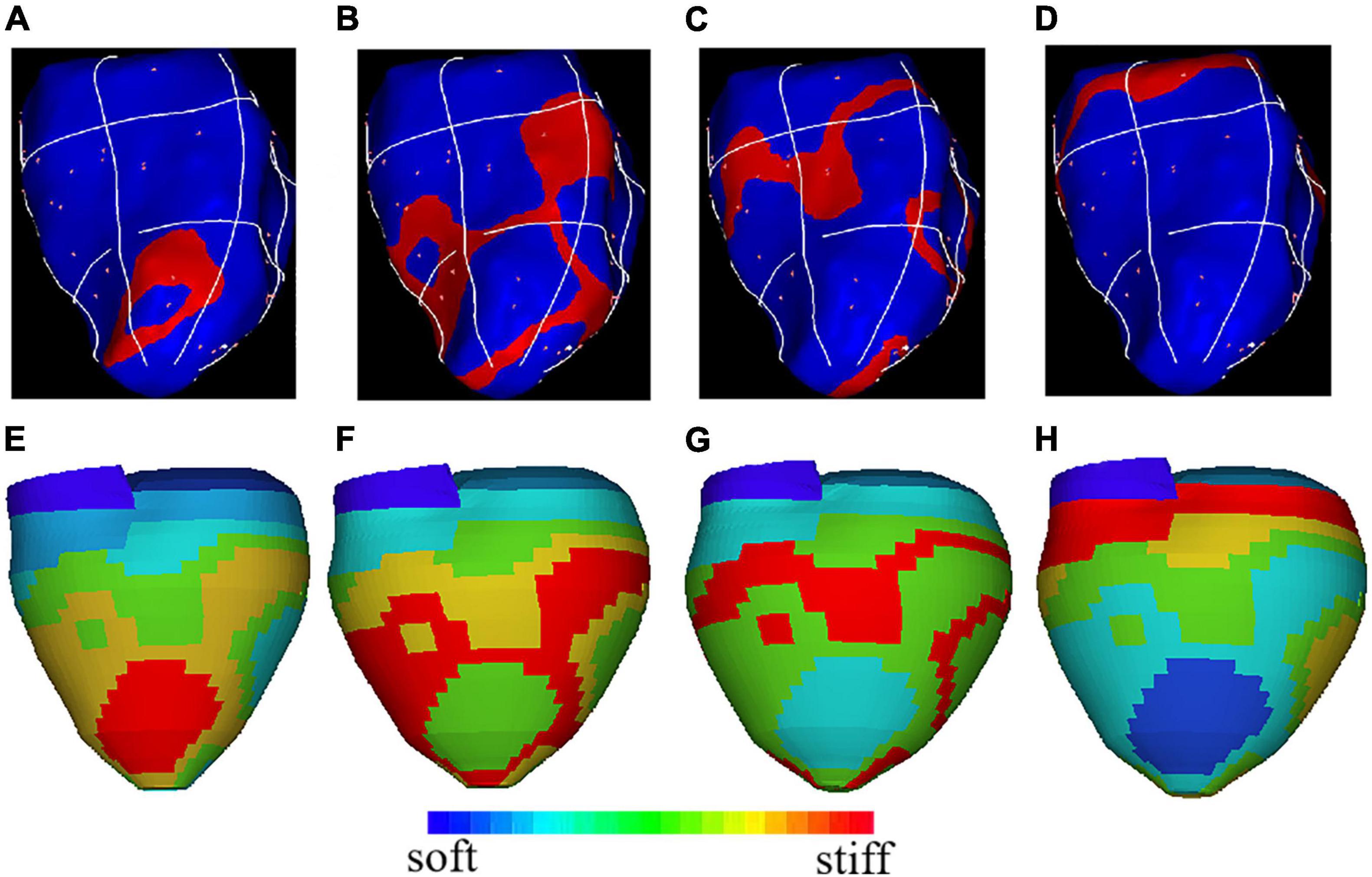
Frontiers Optimization of Left Ventricle Pace Maker Location Using Echo-Based Fluid-Structure Interaction Models

Computed tomography validated right ventricular mid‐septal lead implantation using right ventricular angiography - Shenthar - 2021 - Journal of Arrhythmia - Wiley Online Library

Pacemaker Lead-Induced Severe Tricuspid Valve Stenosis

