Figure 3 from Stimulated Emission Depletion (STED) Microscopy: from Theory to Practice
Por um escritor misterioso
Descrição
Figure 3. Fluorescence depletion of two common dyes in STED microscopy, Atto647N (black, diamonds) and Atto655 (red, circles), as a function of the depletion laser intensity. Error bars for Atto647N appear smaller than the point size of the average value. - "Stimulated Emission Depletion (STED) Microscopy: from Theory to Practice"

Fluorescence microscopy with diffraction resolution barrier broken

Ultralow power demand in fluorescence nanoscopy with digitally

Stimulated Emission Depletion Microscopy

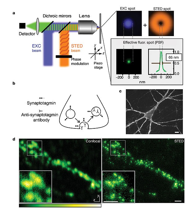
STED microscopy reveals that synaptotagmin remains clustered after

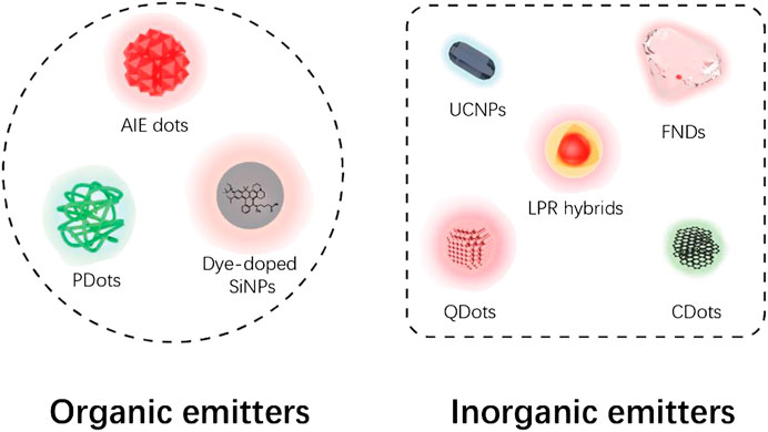
Frontiers Shedding New Lights Into STED Microscopy: Emerging

Nanoparticle-Assisted Stimulated-Emission-Depletion Nanoscopy

Ultralow power demand in fluorescence nanoscopy with digitally

Stimulated Emission Depletion Lithography with Mercapto-Functional
Time-gating improves the spatial resolution of STED microscopy


