Morphology of Leydig cells in the testes after in vivo MCP-1 treatment.
Por um escritor misterioso
Descrição

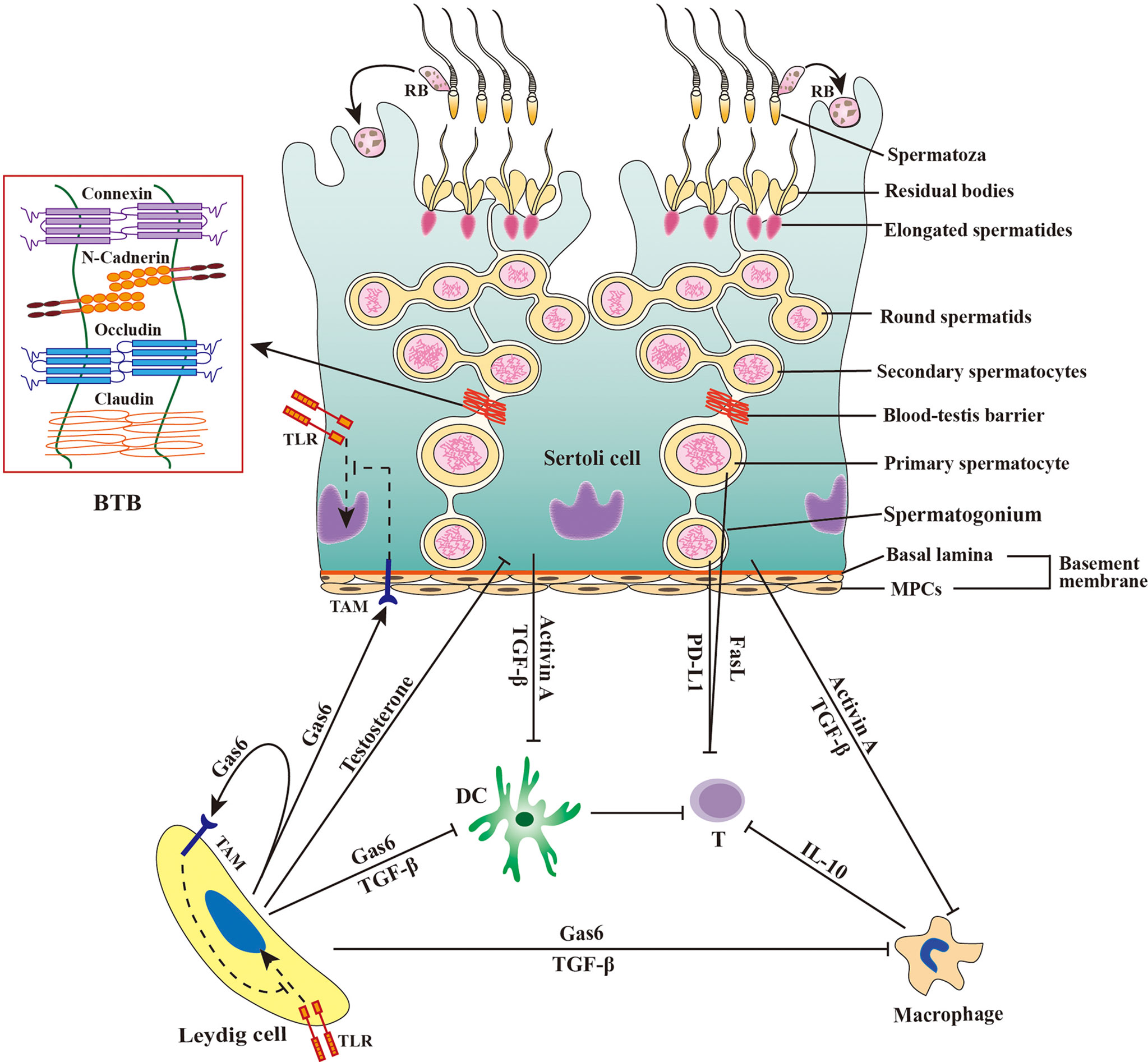
The Sertoli cell: one hundred fifty years of beauty and plasticity - França - 2016 - Andrology - Wiley Online Library

Morphology of Leydig cells in the testes after in vivo PTHrP
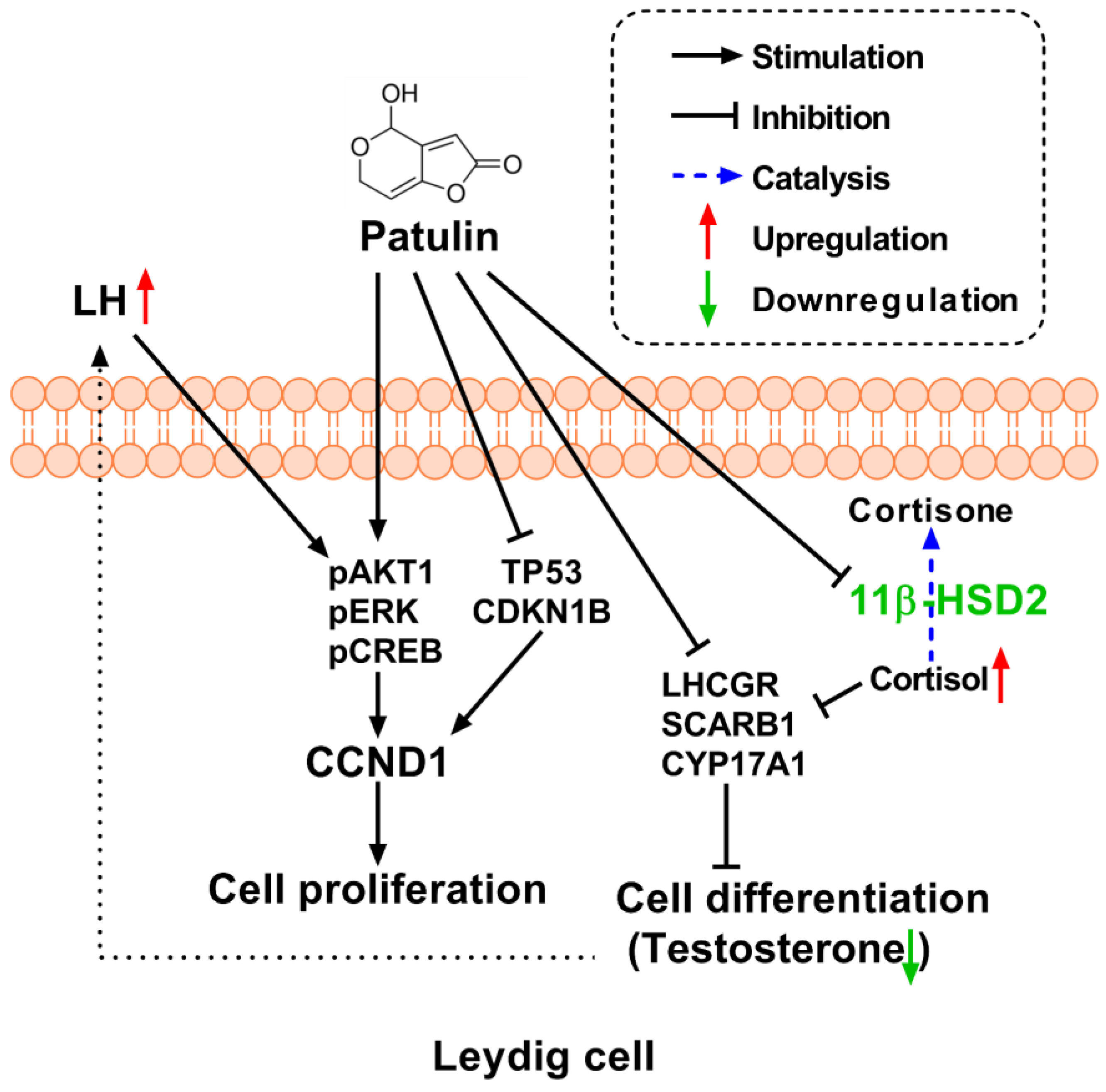
Toxins, Free Full-Text

Morphology of Leydig cells in the testes after in vivo MCP-1 treatment.
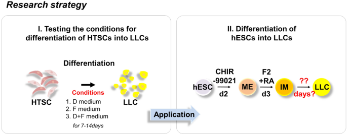
Rapid Differentiation of Human Embryonic Stem Cells into Testosterone-Producing Leydig Cell-Like Cells In vitro

Effect of TNF on testosterone production. Leydig cells were cultured in
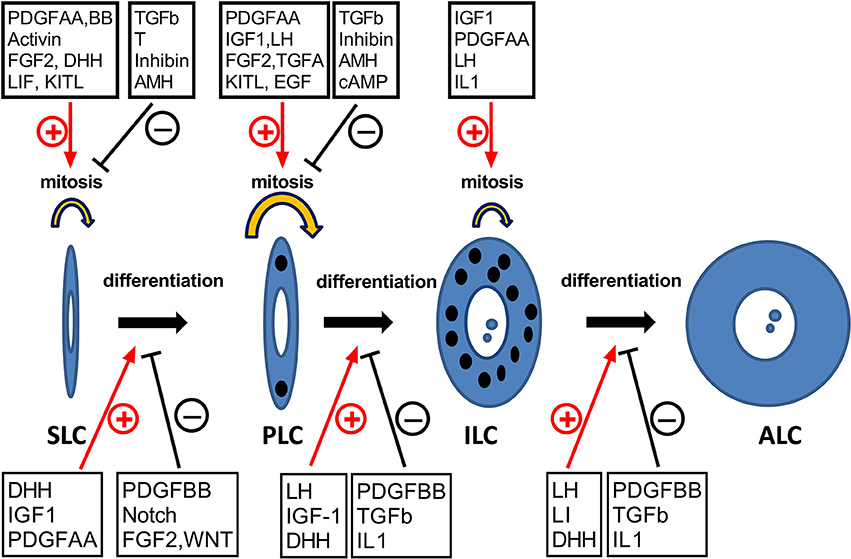
Frontiers Insights into the Development of the Adult Leydig Cell Lineage from Stem Leydig Cells

Therapeutic application of Sertoli cells for treatment of various diseases - ScienceDirect
Frontiers Viral tropism for the testis and sexual transmission

Morphology of Leydig cells in the testes after in vivo MCP-1 treatment.