Figure 1. [The normal human retina fundus]. - Webvision - NCBI
Por um escritor misterioso
Descrição
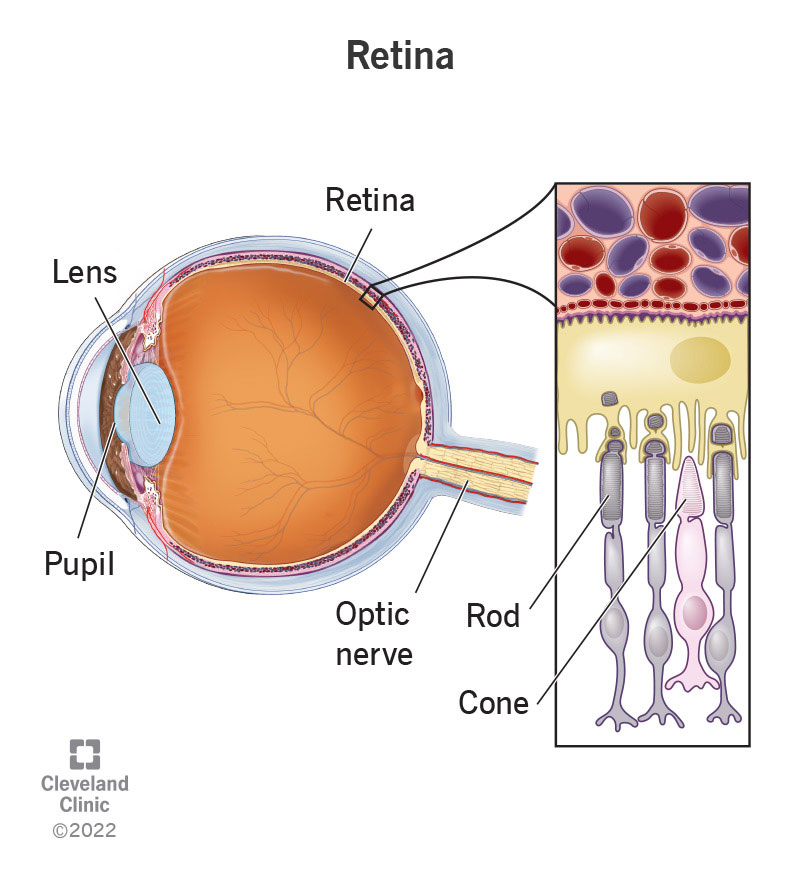
The normal human retina fundus photo shows the optic nerve (right), blood vessels and the position of the fovea (center).
![Figure 1. [The normal human retina fundus]. - Webvision - NCBI](https://www.ncbi.nlm.nih.gov/books/NBK11553/bin/clinicalergf24.jpg)
Figure 24, [Fundus photo and bright-flash ERG of patient with retinoschisis.]. - Webvision - NCBI Bookshelf
![Figure 1. [The normal human retina fundus]. - Webvision - NCBI](https://www.mdpi.com/cells/cells-12-01987/article_deploy/html/images/cells-12-01987-g001.png)
Cells, Free Full-Text
![Figure 1. [The normal human retina fundus]. - Webvision - NCBI](https://media.springernature.com/full/springer-static/image/art%3A10.1038%2Fs41467-019-12917-9/MediaObjects/41467_2019_12917_Fig1_HTML.png)
Single-nuclei RNA-seq on human retinal tissue provides improved transcriptome profiling
![Figure 1. [The normal human retina fundus]. - Webvision - NCBI](https://onlinelibrary.wiley.com/cms/asset/0e5a34c5-0d5d-4cf7-b312-567d11a45ab7/aos15226-fig-0001-m.jpg)
Lipid metabolism and retinal diseases - Gabrielle - 2022 - Acta Ophthalmologica - Wiley Online Library
![Figure 1. [The normal human retina fundus]. - Webvision - NCBI](https://media.springernature.com/full/springer-static/image/art%3A10.1038%2Fgim.2014.95/MediaObjects/41436_2015_Article_BFgim201495_Fig1_HTML.jpg)
Early-onset autosomal recessive cerebellar ataxia associated with retinal dystrophy: new human hotfoot phenotype caused by homozygous GRID2 deletion
![Figure 1. [The normal human retina fundus]. - Webvision - NCBI](https://www.ncbi.nlm.nih.gov/books/NBK554706/bin/Archetecture_Fovea-Image012.gif)
The Architecture of the Human Fovea - Webvision - NCBI Bookshelf
![Figure 1. [The normal human retina fundus]. - Webvision - NCBI](http://webvision.med.utah.edu/wp-content/uploads/2018/05/sagschem.jpg)
Simple Anatomy of the Retina by Helga Kolb – Webvision
![Figure 1. [The normal human retina fundus]. - Webvision - NCBI](https://www.researchgate.net/publication/242466981/figure/fig1/AS:298496365219842@1448178487460/A-Normal-fundus-of-OD-B-Fundus-of-OS-showing-foveal-retinal-pigment-epithelial.png)
A) Normal fundus of OD; (B) Fundus of OS showing foveal retinal
![Figure 1. [The normal human retina fundus]. - Webvision - NCBI](http://webvision.instead-technologies.com/wp-content/uploads/2014/06/nervefibershuman1.jpg)
1.2 Simple Anatomy of the Retina. By Helga Kolb – Webvision
![Figure 1. [The normal human retina fundus]. - Webvision - NCBI](https://www.ncbi.nlm.nih.gov/books/NBK11533/bin/sretinaf16.gif)
Simple Anatomy of the Retina - Webvision - NCBI Bookshelf